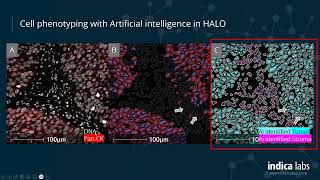
Discovery Notes: Presented by Dr Metta Jana from the Centre for Advanced Histology & Microscopy at the Peter MacCallum Cancer Centre. With its advanced deep learning networks and streamlined train-by-example workflow,

Halo Image Analysis - Comparison Points

This practical guide collects Halo Image Analysis through meaning, examples, related intent, useful checks, and follow-up paths without locking every page into the same repeated structure.

In addition, this page also connects Halo Image Analysis with for broader topic coverage.

Comparison Points

Presented by Dr Metta Jana from the Centre for Advanced Histology & Microscopy at the Peter MacCallum Cancer Centre. With its advanced deep learning networks and streamlined train-by-example workflow,

Information Where It Fits

This part keeps Halo Image Analysis connected to practical references instead of leaving it as a single isolated phrase.

General User-Friendly Overview

Halo Image Analysis can be reviewed through a clear overview first, then compared with related entries and supporting context.

Context Useful Tips

Use the related entries as follow-up paths when you need more examples, current details, or alternative wording.

Relevant points collected here

  • With its advanced deep learning networks and streamlined train-by-example workflow,
  • Presented by Dr Metta Jana from the Centre for Advanced Histology & Microscopy at the Peter MacCallum Cancer Centre.

Why this overview helps

Readers often search for Halo Image Analysis because they want a quick explanation, related examples, and practical next steps.

Sponsored

Questions People Also Check

What questions should readers ask about Halo Image Analysis?

Check freshness, source quality, related examples, and any requirements or limitations before relying on one answer.

What should be checked first?

Readers should check the main context, important requirements, source freshness, and any details that may change over time.

What should readers do next?

Readers can review the linked topics, compare several sources, and verify important details before acting on the information.

How can readers narrow down Halo Image Analysis?

Readers can narrow it by adding location, year, product name, provider, price range, purpose, or the exact problem they want to solve.

Related Visuals

HALO® Image Analysis
Getting Started with Multiplex Fluorescence Image Analysis in HALO®
Bench-to-Bedside Transition of Novel HALO® Assays into Clinical Trials with HALO AP®
Talk 4: Using Halo to analyse OPAL mIHC images
Cell DIVE a robust platform for multiplexed whole slide imaging and single cell analysis
Gaining key phenotypic data from the TME using 8-plex immunofluorescence staining and image analysis
MPIC Tissue Quantification (HALO)
Indica Labs' Integrated HALO® Platforms
HALO AI: Trainable AI for Digital Pathology
122 - Normalizing H&E images and digitally separating Hematoxylin and Eosin components
Sponsored
Read Clear Overview
HALO® Image Analysis

HALO® Image Analysis

Read more details and related context about HALO® Image Analysis.

Getting Started with Multiplex Fluorescence Image Analysis in HALO®

Getting Started with Multiplex Fluorescence Image Analysis in HALO®

Getting Started with Multiplex Fluorescence Image Analysis in HALO®

Bench-to-Bedside Transition of Novel HALO® Assays into Clinical Trials with HALO AP®

Bench-to-Bedside Transition of Novel HALO® Assays into Clinical Trials with HALO AP®

Read more details and related context about Bench-to-Bedside Transition of Novel HALO® Assays into Clinical Trials with HALO AP®.

Talk 4: Using Halo to analyse OPAL mIHC images

Talk 4: Using Halo to analyse OPAL mIHC images

Presented by Dr Metta Jana from the Centre for Advanced Histology & Microscopy at the Peter MacCallum Cancer Centre.

Cell DIVE a robust platform for multiplexed whole slide imaging and single cell analysis

Cell DIVE a robust platform for multiplexed whole slide imaging and single cell analysis

Read more details and related context about Cell DIVE a robust platform for multiplexed whole slide imaging and single cell analysis.

Gaining key phenotypic data from the TME using 8-plex immunofluorescence staining and image analysis

Gaining key phenotypic data from the TME using 8-plex immunofluorescence staining and image analysis

Broadcast via The Pathologist, November 2020. Multiplex staining and

MPIC Tissue Quantification (HALO)

MPIC Tissue Quantification (HALO)

Read more details and related context about MPIC Tissue Quantification (HALO).

Indica Labs' Integrated HALO® Platforms

Indica Labs' Integrated HALO® Platforms

Read more details and related context about Indica Labs' Integrated HALO® Platforms.

HALO AI: Trainable AI for Digital Pathology

HALO AI: Trainable AI for Digital Pathology

With its advanced deep learning networks and streamlined train-by-example workflow,

122 - Normalizing H&E images and digitally separating Hematoxylin and Eosin components

122 - Normalizing H&E images and digitally separating Hematoxylin and Eosin components

Read more details and related context about 122 - Normalizing H&E images and digitally separating Hematoxylin and Eosin components.